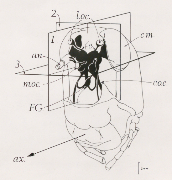
FIGURE 4.1
Diagram illustrating the position of the brain (black) within the head of Schistocerca gregaria.
Key:
cm. compound eye
an. antenna
m.oc. median ocellus
l.oc. lateral ocellus
F.G. frontal ganglion
ax. axis of the ventral nerve cord
The three main planes of section are shown : 1 = longitudinal, 2 = transverse and 3 = horizontal.
Original drawing by Les Williams